1. Basic introduction: Different types of pigments usually appear in both normal and abnormal liver cytology. Because most pigments are in the liver cells and are green with Reischer Gimssa or Dev staining, special stains are often used to distingui...
1. Basic introduction:
Different types of pigments usually appear in both normal and abnormal liver cytology. Because most pigments are in the liver cells and are green with Reischer Gimssa or Dev staining, special stains are often used to distinguish different types of pigments.
1. Lipofuscin:
In normal hepatocytes, most of the pigments are lipofuscin, a normal "loss" pigment, which is related to indigestive substances accumulated in the lysosome. Many granular dark green pigments are visible in the liver cells of elderly dogs and cats. The accumulation of lipofuscin is considered to be part of normal aging and cannot directly represent disease. A modified acid-resistant dye (Ziehl-Neelsen) was used to confirm the presence or absence of lipofuscin.
2. Bile:
The accumulation of bile in hepatocytes is manifested as yellow to oily green or dark green to black pigments of varying sizes. Excessive bilirubin, especially when a bile-filled tube plug appears, suggests cholestasis, which may be a precursor to clinical jaundice or hyperbilirubinemia. In cytologic samples, this accumulation manifests as a unique, intact and broken dark green, yellow or olive green green tubular accumulation. Hall’s bile stain can be used to confirm the presence of bile (bilirubin). Causes of bile obstruction, such as liver lipid deposition or tumor cell infiltration, may be observed in cytology samples, although in many cases the cause is undeterminable.
3. Copper:
Stained with Diff or Reischer Gimsa, copper may be seen in the liver, which manifests as rough, granular, light blue, fragmented pigments, when present in large quantities, although special staining is required, such as red amine. Excessive copper may cause liver damage or necrosis and accompany inflammation. Copper accumulation may be a primary disease, such as hereditary diseases in Bedlington thick-haired dogs, or secondary changes in chronic hepatitis and other liver diseases.
4. Hemosiderin:
Stained with Diff or Reischer Gimssa, the iron-containing yellow-brown to blue-black pigment appears in the liver cytoplasm. The clear diagnosis requires special staining, and Tuprus blue staining. Methostein is a kind of iron-containing pigment particles that accumulate in liver cells. In diseased states, such as hemolytic anemia and chronic inflammation, it can also occur when the diseased animal is subjected to repeated blood perfusion or iron injection.
2. Cytology:
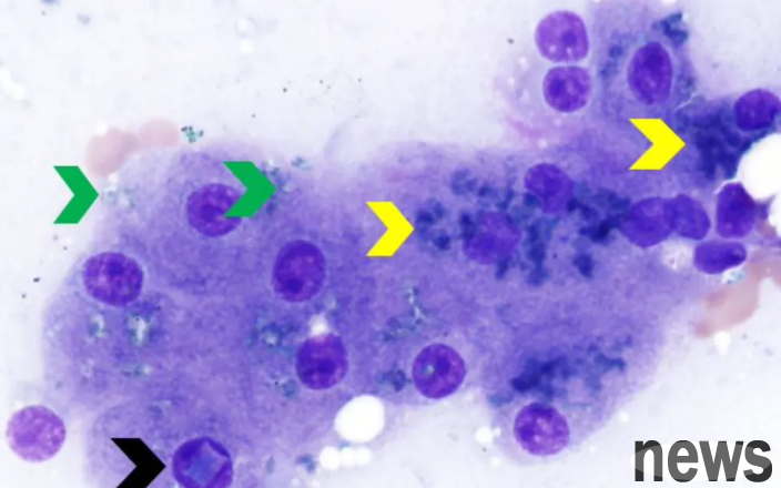
Cancerology, Diff 1000X: Note that the granular black-green pigment in liver cells is lipofuscin (yellow arrow); the black arrow refers to the rectangular inclusion body in the nucleus, occasionally, and the clinical significance is unknown; a small amount of light blue, slightly fragmented pigment in liver cells is confirmed to be copper (green arrow) through erythramine staining.
Cat liver cytology, Reis Gimssa 1000X: The large amount of granular dark green pigment referred to by the yellow arrow is lipofuscin, from clinically normal elderly cats.
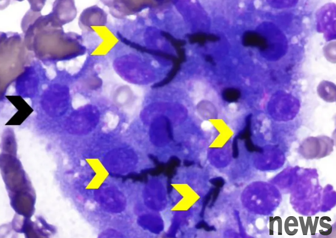
Cytology of cholestasis in canine liver, Reischer Gimssa 1000X: Note clustered hepatocytes (black arrows) mixed with tubular plugs filled with dark green to black bile (yellow arrows).
Also for canine liver cholestasis cytology, Diff 500X: The yellow arrow confirms that several small tube plugs filled with bile, yellow to oily green.

Hepatic cytology in dogs with chronic hemolytic anemia, Reich staining 1000X: Hemosiderin-containing pigment appears as a brown-yellow pigment in hepatocytes, and its surrounding red blood cell lines and myeloid cell line precursor cells suggest the presence of extramedullary hematopoiesis in the liver.